What is iCAT 3-D Imaging?
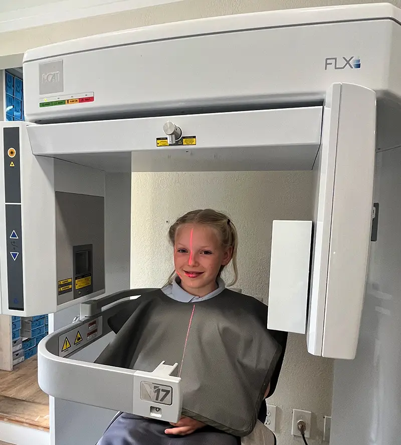
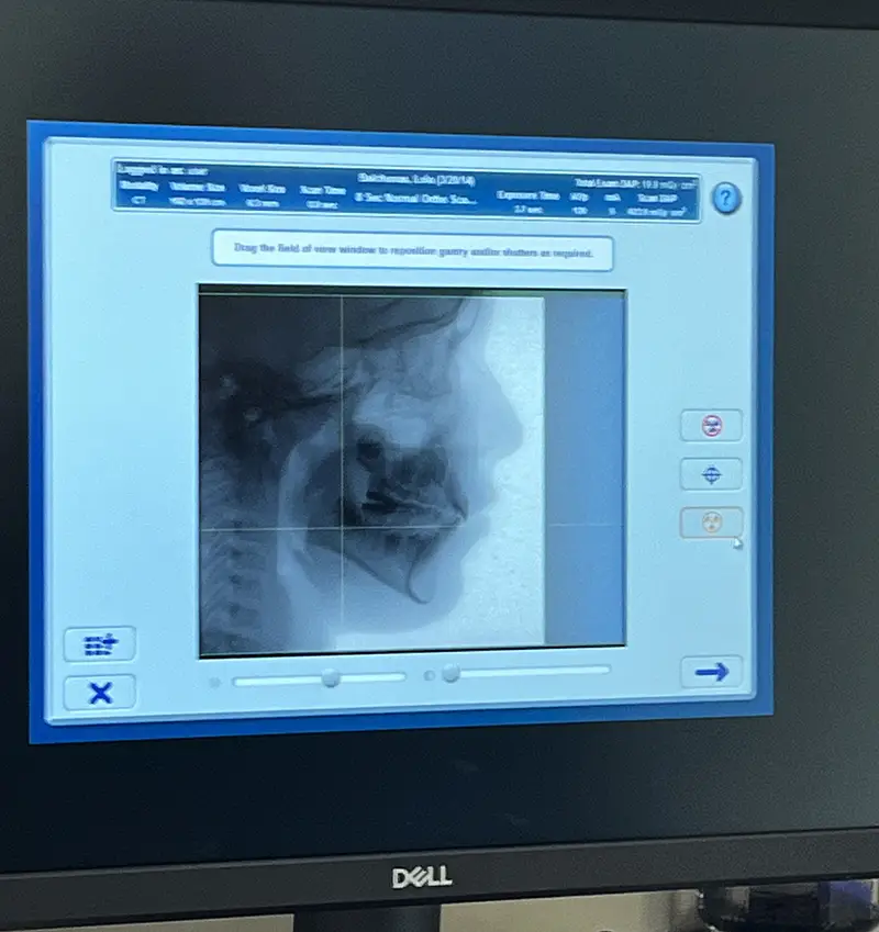
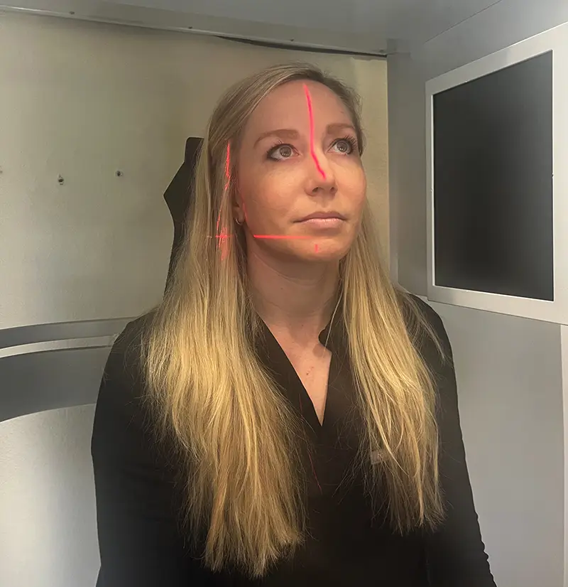
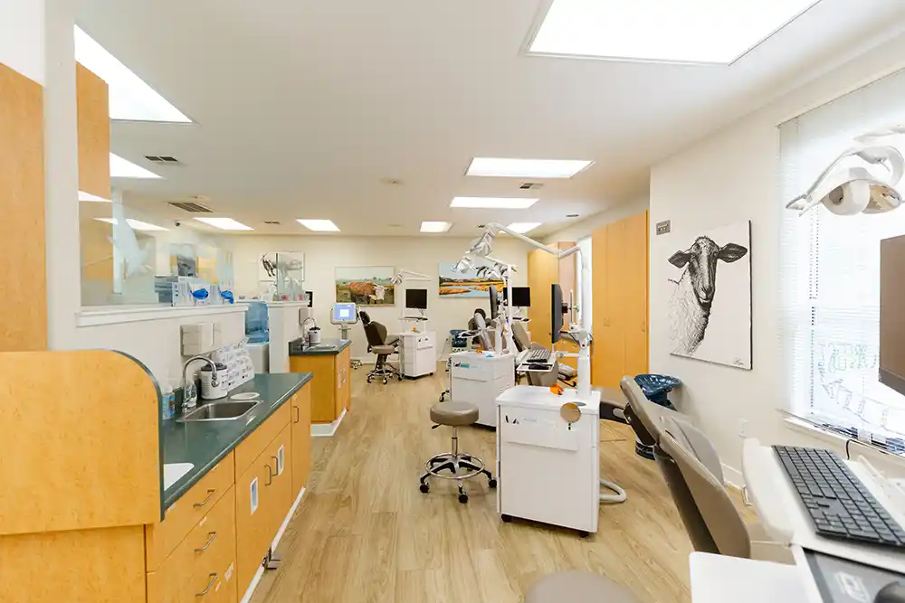
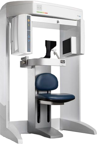
At Bales & King Orthodontics we are pleased to offer 3D imaging for each new patient. This low dose x ray is called a CBCT (cone-beam computed technology) and provides us with much more information than traditional 2D imaging. Traditional 2-D scans give orthodontists an approximation of a person’s teeth, jaw and skull. The detailed, 360º picture provided by the i-CAT system, on the other hand, enables us to completely visualize and evaluate the erupted and unerupted teeth, jaws, jaw joints, and airways of each patient and plan their orthodontic treatment appropriately. The effective dose is the same as the background radiation from spending a few days outside in the sunshine. In fact, the dose is less than that of traditional 2D panoramic and cephalometric imaging.
Our 3D CBCT scan is an invaluable tool in screening for sleep related breathing disorders.
It allows us to visualize enlarged tissues such as the tonsils and turbinates, as well as congested areas, such as the sinuses, that may be impeding the flow of air during sleeping. Orthodontists are also trained to detect oral manifestations of labored breathing during sleep such as narrow dental arches, dental crossbites, open bites, tongue thrusts, etc. The iCAT imaging system allow us to capture 3D x-rays to analyze teeth, roots, jaw joints, skeletal balance, airway, and sinuses without magnification or distortion. The scan takes less than 10 seconds and shows significantly more information to allow accurate diagnosis and unprecedented treatment planning ability.
What Can Be Detected on CBCT?
- Impacted teeth and precise location. (something not possible with 2D imaging)
- Precise development path of adult teeth behind the baby teeth, including ectopic teeth
- Supernumerary teeth that can often go undetected with 2D imaging
- Ankylosed teeth
- Genetically missing teeth
- Periodontal condition including bone levels and position of roots inside of the jaw bone
- Skeletal asymmetries and jaw growth imbalances
- Transverse discrepancies of the upper and lower jaws
- Nasal structures: Turbinate enlargement, septum deviations, concha bullosa, nasal stenosis, space inside of the nose for breathing
- Sinuses: We can visualize sinus occlusion as well as how close the roots of the teeth are to the sinuses
- Enlarged tonsils and adenoids
- Tongue position and soft palate morphology
- Airway measurements to help us determine where possible airway obstruction exist
- Jaw Joints: Can visualize the TMJ and detect if any abnormal joint space, remodeling and possible signs of degeneration exist
